27 October 2026
Why Is the Extracellular Matrix (ECM) Essential to Regenerative Aesthetics?
Understanding Skin Aging, Collagen Biostimulation, and Tissue Remodeling Collagen has long been a central focus in aesthetic medicine. However, modern skin biology suggests that skin aging involves more than collagen loss. It also involves progressive changes in the extracellular matrix (ECM), the biological environment surrounding cells.
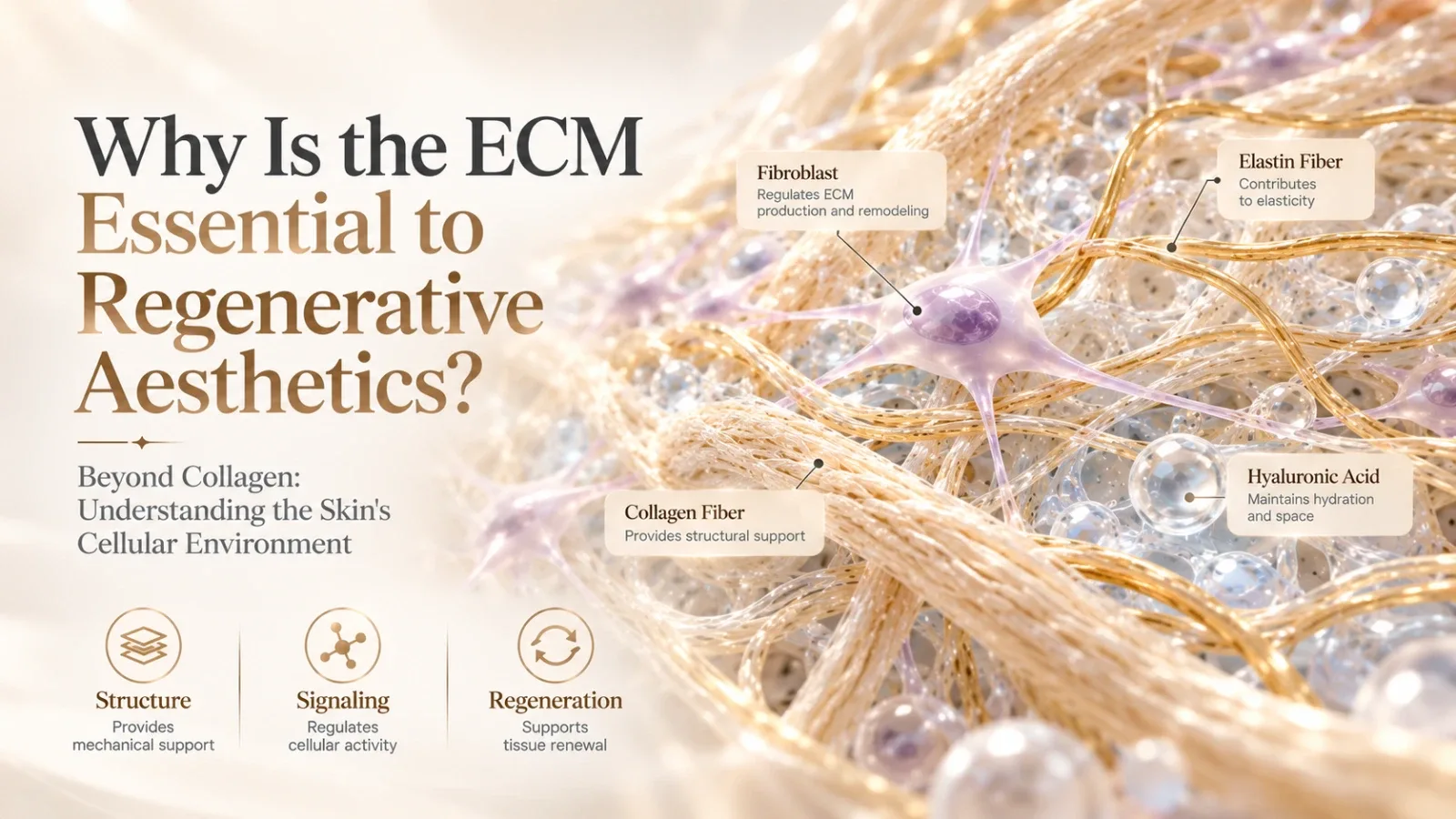
As ECM integrity declines, fibroblast function may also change, influencing collagen synthesis and degradation. Understanding this relationship has become increasingly important in the study of skin aging and regenerative biomaterials.
1. The ECM Is More Than a Structural Framework
The extracellular matrix is a complex three-dimensional network composed of collagen, elastin, hyaluronic acid, proteoglycans, and other structural molecules.
Within the dermis, the ECM provides mechanical strength, elasticity, and hydration support. It also interacts with cellular receptors, including integrins, influencing cell adhesion, migration, proliferation, and tissue repair.
Hynes and Naba emphasized that ECM proteins and associated molecules participate in a wide range of cellular functions rather than serving solely as passive structural components.[1]
The ECM is therefore both a structural environment and an important regulator of cellular behavior.
2. How Does ECM Deterioration Contribute to Skin Aging?
As skin ages, collagen synthesis gradually decreases, while matrix metalloproteinases (MMPs) contribute to collagen degradation and fragmentation.
These changes weaken the structural integrity of the ECM and affect fibroblast interactions with surrounding collagen fibers.
Research has shown that fragmented collagen can impair fibroblast spreading and mechanical tension, reducing collagen production while potentially increasing the expression of matrix-degrading enzymes.[2][3]
This creates a progressive cycle:
Collagen fragmentation → ECM disruption → Altered fibroblast function → Reduced collagen synthesis → Further ECM deterioration
Skin aging is therefore not simply a loss of structural proteins. It also involves changes in the relationship between cells and their surrounding microenvironment.
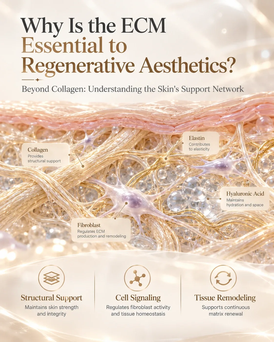
3. Mechanotransduction: How Does the ECM Influence Fibroblasts?
Fibroblasts continuously respond to the mechanical characteristics of their environment, including matrix stiffness, elasticity, and tension.
This process is known as mechanotransduction.
A landmark study published in Science in 2005 demonstrated that cells can sense substrate stiffness and adjust their behavior through cellular adhesion and cytoskeletal mechanisms.[4]
In skin tissue, changes in ECM architecture can therefore influence the mechanical signals received by fibroblasts.
As collagen fibers become fragmented and the matrix loses structural integrity, fibroblast–matrix interactions may weaken, affecting collagen homeostasis.
This helps explain why regenerative aesthetics increasingly considers not only collagen quantity, but also matrix organization, cellular interaction, and long-term tissue function.
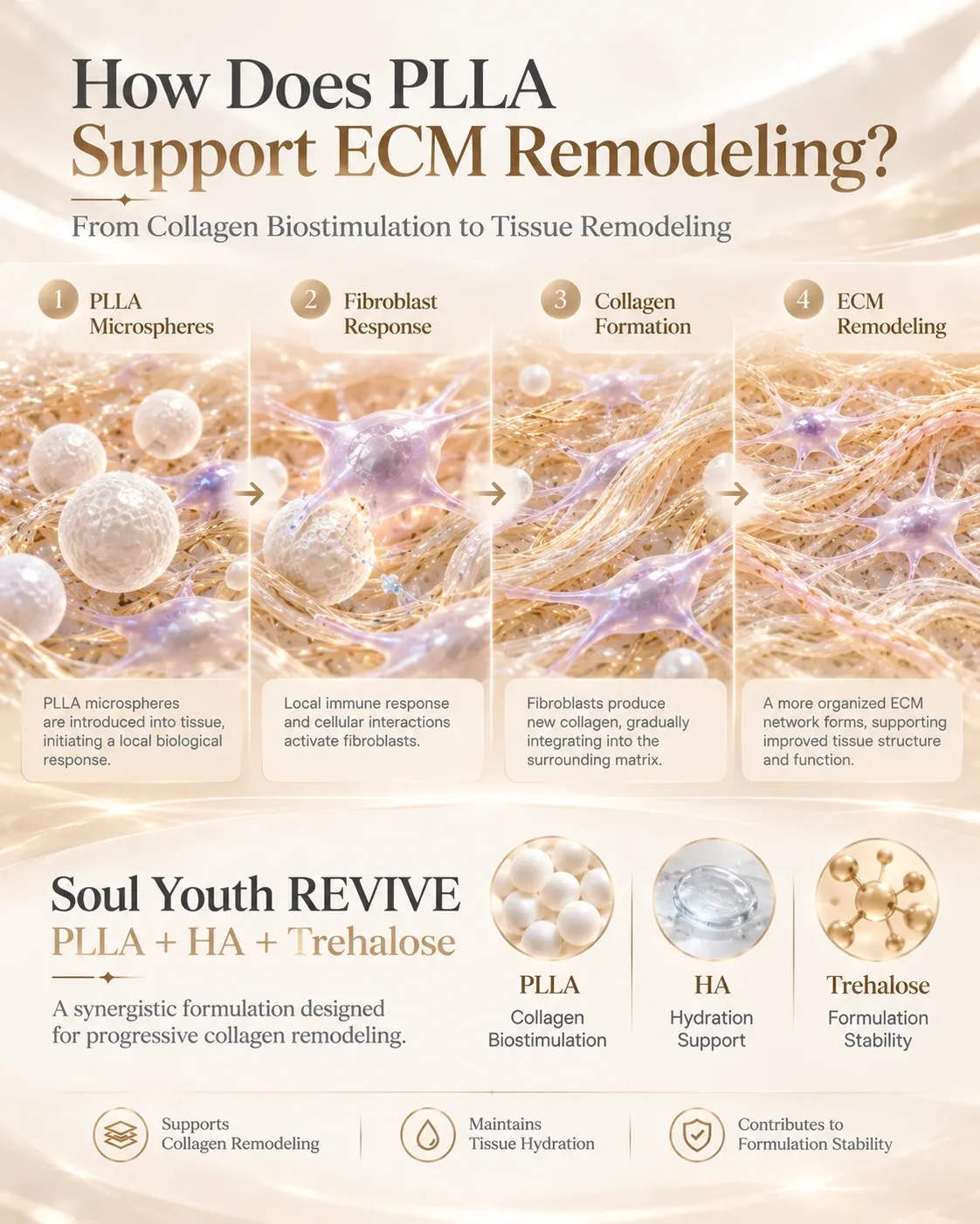
4. How Does PLLA Support Collagen Biostimulation and ECM Remodeling?
Poly-L-Lactic Acid (PLLA) is a representative collagen biostimulatory material.
Rather than directly replacing lost collagen, PLLA microspheres initiate a local material–tissue response that supports progressive collagen formation.
Following implantation, immune cells participate in the recognition and gradual degradation of the material. Through subsequent biological signaling, fibroblasts contribute to new collagen synthesis and local ECM remodeling.
Laboratory studies have provided further insight into this mechanism. An in vitro study found that PLLA increased Type I collagen-related gene expression and procollagen production in human dermal fibroblasts through pathways associated with TGF-β/Smad signaling.[5]
These findings help explain PLLA's biostimulatory potential. However, collagen stimulation should not be interpreted as complete restoration of all ECM structures or functions.
Clinical outcomes depend on material characteristics, microsphere design, injection technique, and individual tissue responses.
5. From ECM Science to Soul Youth Regenerative Solutions
Understanding ECM biology provides an important foundation for regenerative biomaterial development.
Soul Youth REVIVE combines PLLA + HA + Trehalose within an integrated formulation strategy.
PLLA acts as the primary collagen biostimulatory component, supporting progressive collagen formation through material–tissue interactions. HA contributes hydration and volume support, while Trehalose plays a role in formulation stability.
Microsphere morphology, particle distribution, and formulation consistency are also important considerations in regenerative biomaterial design.
This reflects a broader direction in regenerative aesthetics: moving beyond individual ingredients toward more integrated material engineering.
At the same time, product-specific clinical evidence remains essential when evaluating the safety and effectiveness of any regenerative aesthetic formulation.
To learn more about how these material design principles are reflected in our formulation approach, explore Soul Youth REVIVE, a PLLA-based collagen biostimulator combining HA and Trehalose.
For more information on composition, product characteristics, and professional applications, visit our REVIVE Knowledge Centre.
Conclusion: Regenerative Aesthetics Goes Beyond Collagen
The extracellular matrix connects tissue architecture, cellular behavior, and biological signaling.
Understanding how ECM structure influences fibroblast activity—and how fibroblasts participate in matrix renewal—provides a more comprehensive perspective on regenerative biomaterials.
The future of regenerative aesthetics is not simply about stimulating more collagen. It is about understanding how biomaterial design and tissue interactions can support a healthier, more functional extracellular environment.
References
[1] Hynes RO, Naba A. (2012).
Overview of the Matrisome—An Inventory of Extracellular Matrix Constituents and Functions.
Cold Spring Harbor Perspectives in Biology, 4(1), a004903.
DOI: 10.1101/cshperspect.a004903
[2] Quan T, Fisher GJ. (2015).
Role of Age-Associated Alterations of the Dermal Extracellular Matrix Microenvironment in Human Skin Aging: A Mini-Review.
Gerontology, 61(5), 427–434.
DOI: 10.1159/000371708
[3] Fisher GJ, Varani J, Voorhees JJ. (2008).
Looking Older: Fibroblast Collapse and Therapeutic Implications.
Archives of Dermatology, 144(5), 666–672.
DOI: 10.1001/archderm.144.5.666
[4] Discher DE, Janmey P, Wang YL. (2005).
Tissue Cells Feel and Respond to the Stiffness of Their Substrate.
Science, 310(5751), 1139–1143.
DOI: 10.1126/science.1116995
[5] Poly-L-Lactic Acid Increases Collagen Gene Expression and Synthesis in Cultured Dermal Fibroblast (Hs68) Through the TGF-β/Smad Pathway. (2023).
PubMed PMID: 36575891
Specifications, terms and registration status.
Our team supports licensed clinics, distributors and OEM partners — tell us what your market needs.